JUCM:
ECG Challenge #4
A 31-year-old male presents with weakness.
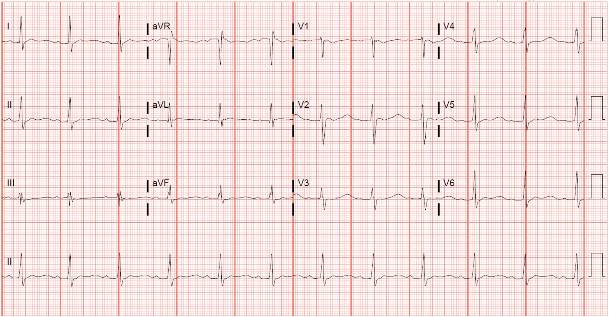
What electrolyte disturbances cause the interval problem seen in this ECG? Which dysrhythmia is this patient at risk for?
Watch Video Summary
What electrolyte disturbances cause the interval problem seen in this ECG?
Hypokalemia, hypomagnesemia, and hypocalcemia can all cause a prolongation of the QT interval. While hypokalemia and hypomagnesemia both delay the repolarization phase (phase 3) of the of the cardiac action potential (creating wide-based T waves, U waves, or a fusion of both), hypocalcemia prolongs the QT interval by way of extending the plateau phase (phase 2) of the cardiac action potential (lengthening the ST segment but with a normal T wave, Figure 1). This ECG is consistent with hypokalemia and/or hypomagnesemia.1
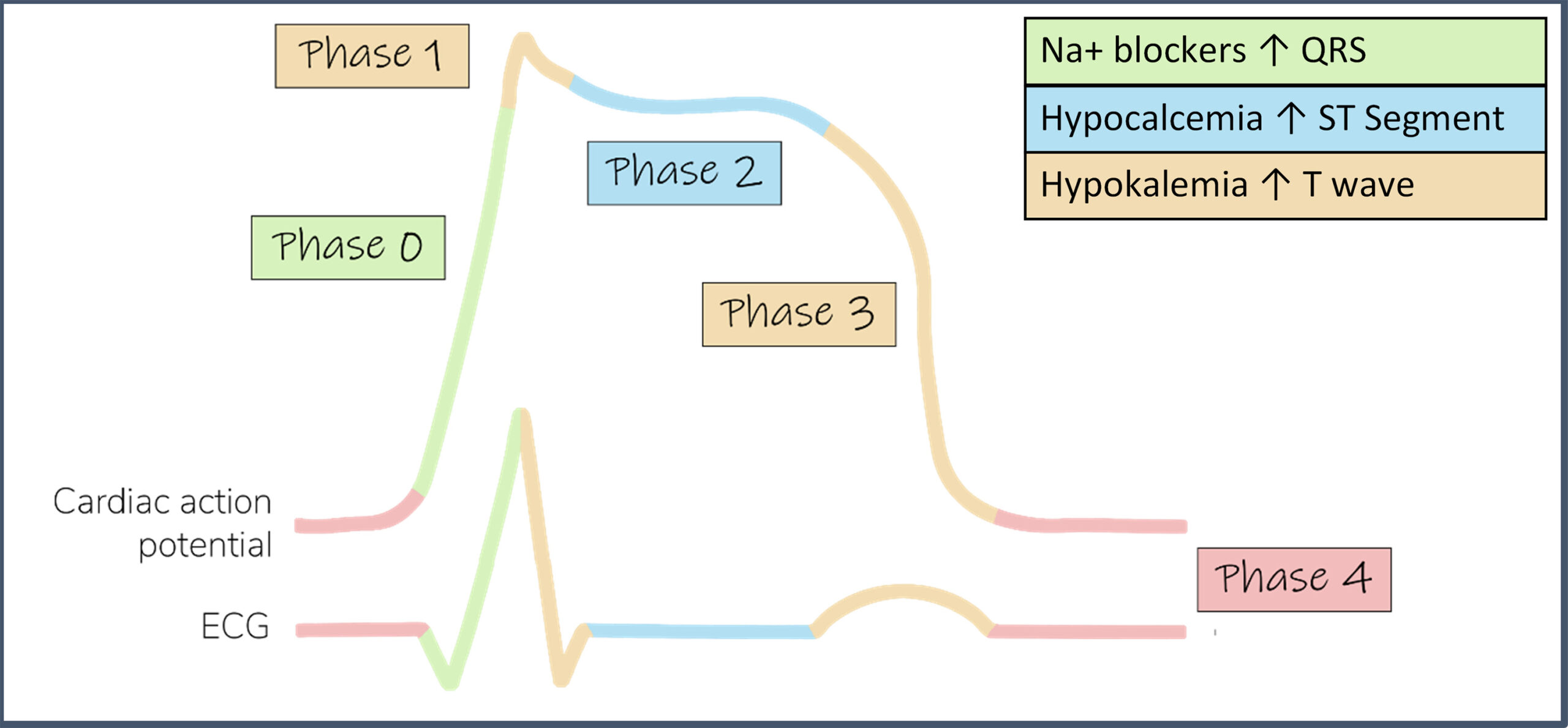
Figure 1. This schematic demonstrates the various phases of the cardiac action potential as they relate to the ECG waveform. In phase 0, fast acting voltage-gated sodium channels open and a rapid influx of sodium results. Sodium channel blocking agents tend to predominantly affect phase 0 and widen the QRS complex. During phase 2, voltage-gated potassium (efflux) and calcium (influx) channels tend to maintain a relative potential plateau. Since phase 2 represents the ST segment, hypocalcemia tends to produce a long ST segment, but a normal T wave. Finally, in phase 3, potassium channels allow more potassium to leak and “repolarize” the cell. Hypokalemia tends to delay this phase, creating broad-based T waves, U waves, and T-U fusions.
Which dysrhythmia is this patient at risk for?
Torsades de Pointes due to early afterdepolarizations/triggered activity during the repolarization phase (R on T phenomenon).2 It’s worth noting that the QT correction formulas (e.g. Bazett’s formula) are not very good outside a normal heart rate range, and that data supporting the use of a QT nomogram is emerging.3,4 Measuring the QT can be challenging, and many different methods exist. One simple method is the “half the RR” rule – the QT interval is prolonged if it occupies more than half the R-R interval. The “half the RR” rule is a conservative estimate at normal and tachycardic rates, but perhaps a better option at bradycardic rates is an absolute cut-off of 485 ms.4
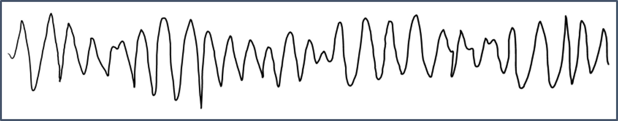
Figure 2. Torsade de Pointes
Pearls for Urgent Care Management
- Symptomatic patients with prolonged QT should be transferred to a higher level of care.
- Common electrolytes that cause prolonged QT include hypokalemia, hypomagnesemia, and hypocalcemia. If able, commence electrolyte repletion prior to transfer.
References
- Diercks DB, Shumaik GM, Harrigan RA, Brady WJ, Chan TC. Electrocardiographic manifestations: Electrolyte abnormalities. Journal of Emergency Medicine. 2004;27(2):153-160. doi:10.1016/j.jemermed.2004.04.006
- Al-Khatib SM, Stevenson WG, Ackerman MJ, et al. 2017 AHA/ACC/HRS Guideline for Management of Patients With Ventricular Arrhythmias and the Prevention of Sudden Cardiac Death. Journal of the American College of Cardiology. 2018;72(14):e91-e220. doi:10.1161/CIR.0000000000000549
- Isbister GK. Risk assessment of drug-induced QT prolongation. Australian prescriber. 2015;38(1):20-24. doi:10.18773/austprescr.2015.003
- Rischall ML, Smith SW, Friedman AB. Screening for QT Prolongation in the Emergency Department: Is There a Better “Rule of Thumb?” Western Journal of Emergency Medicine. 2020;226(2). doi:10.5811/westjem.2019.10.40381