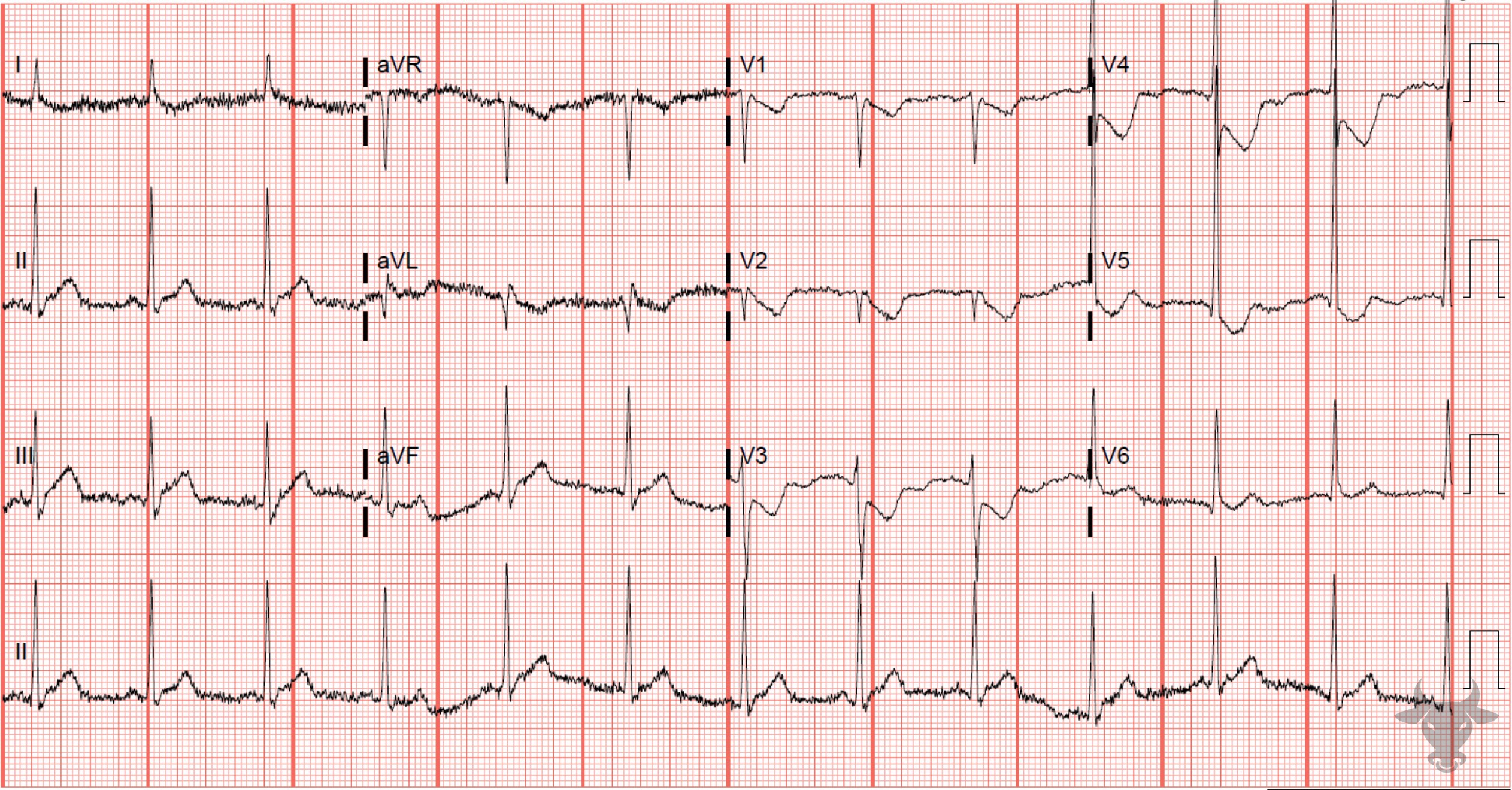
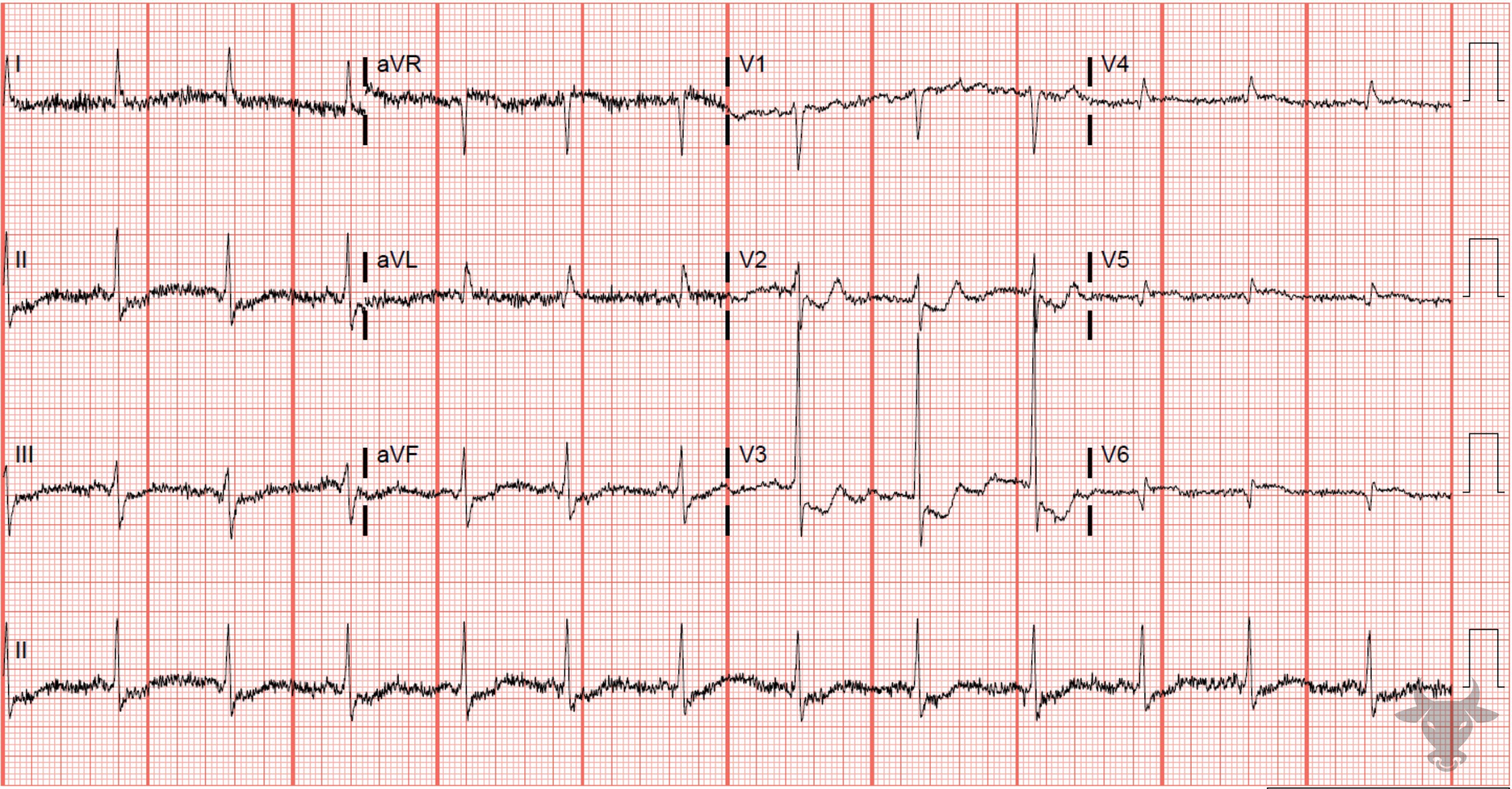
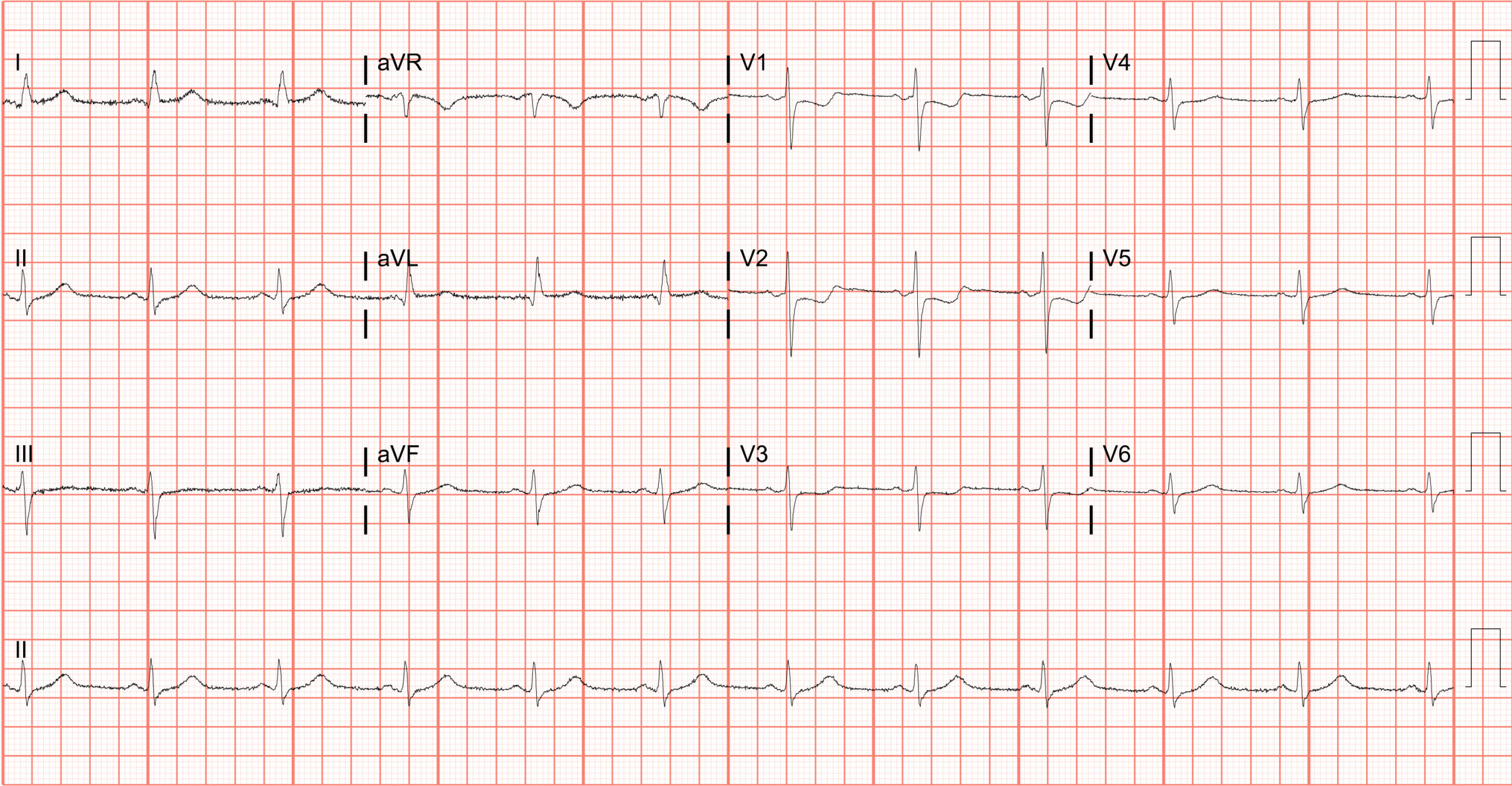
Posterior myocardial infarction is commonly confused with anterior subendocardial ischemia; however, the T waves are expected to be negatively deflected with anterior subendocardial ischemia (unless early on when they can mirror posterior hyperacute T waves). 5-10% of all myocardial infarctions are isolated posterior and not associated with inferior ST elevation. Isolated posterior myocardial infarctions are associated with longer door-to-balloon times and worse outcomes because they are frequently missed. ST elevation of at least 0.5 mm in posterior leads can help secure the diagnosis.
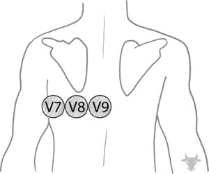
Posterior leads placed under the left scapula and designated as V7, V8, and V9.