Limb Lead Reversal

Between 0.4 and 4% of ECG’s obtained in various settings demonstrate features of lead misplacement. Lead misplacement may either obscure important findings or simulate the appearance of ectopic rhythms, conduction disturbances, chamber abnormalities, and ischemia/infarction – resulting in unnecessary and potentially invasive testing. The key ECG findings suggestive of lead misplacement are dependent on the leads involved. The relationship between limb leads and surface electrodes are defined by Einthoven’s Triangle.


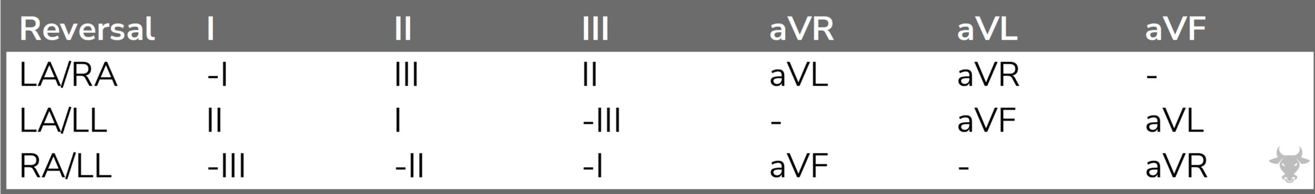
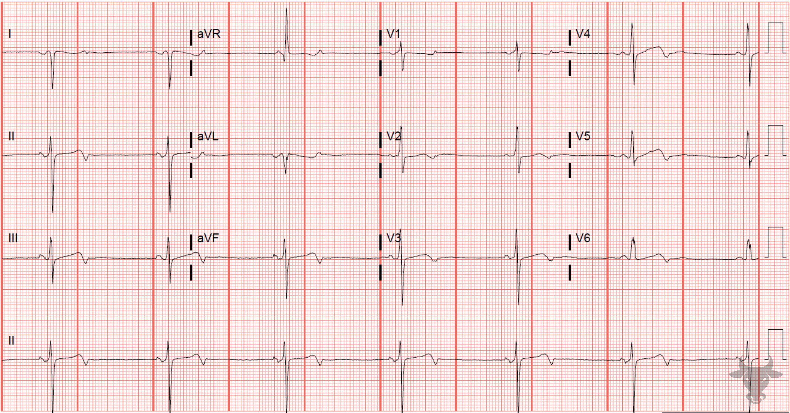
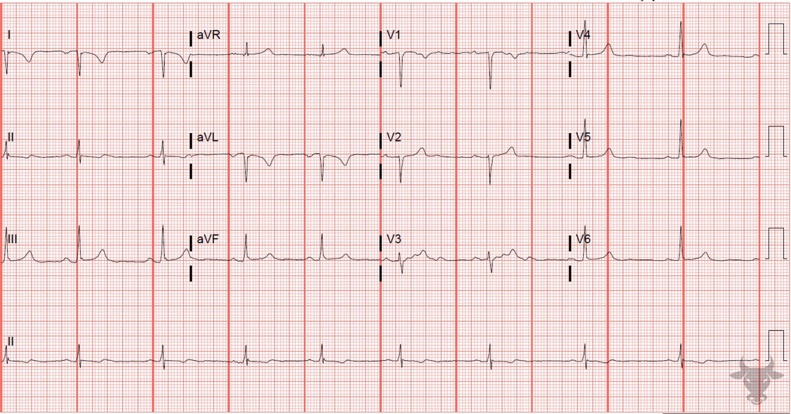
ECG changes resulting from limb lead reversal.

Examples

References

  1. Rudiger, A., Hellermann, J. P., Mukherjee, R., Follath, F. & Turina, J. Electrocardiographic artifacts due to electrode misplacement and their frequency in different clinical settings. Am J Emerg Medicine 25, 174–178 (2007).
  2. Batchvarov, V. N., Malik, M. & Camm, A. J. Incorrect electrode cable connection during electrocardiographic recording. Ep Europace 9, 1081–1090 (2007).
  3. Harrigan, R. A., Chan, T. C. & Brady, W. J. Electrocardiographic Electrode Misplacement, Misconnection, and Artifact. J Emerg Medicine 43, 1038–1044 (2012).
  4. Rosen, A. V., Koppikar, S., Shaw, C. & Baranchuk, A. Common ECG Lead Placement Errors. Part I: Limb lead Reversals. Int J Medical Students 2, 92–98 (2014).
  5. Pérez‐Riera, A. R., Barbosa‐Barros, R., Daminello‐Raimundo, R. & Abreu, L. C. Main artifacts in electrocardiography. Ann Noninvas Electro 23, e12494 (2018).