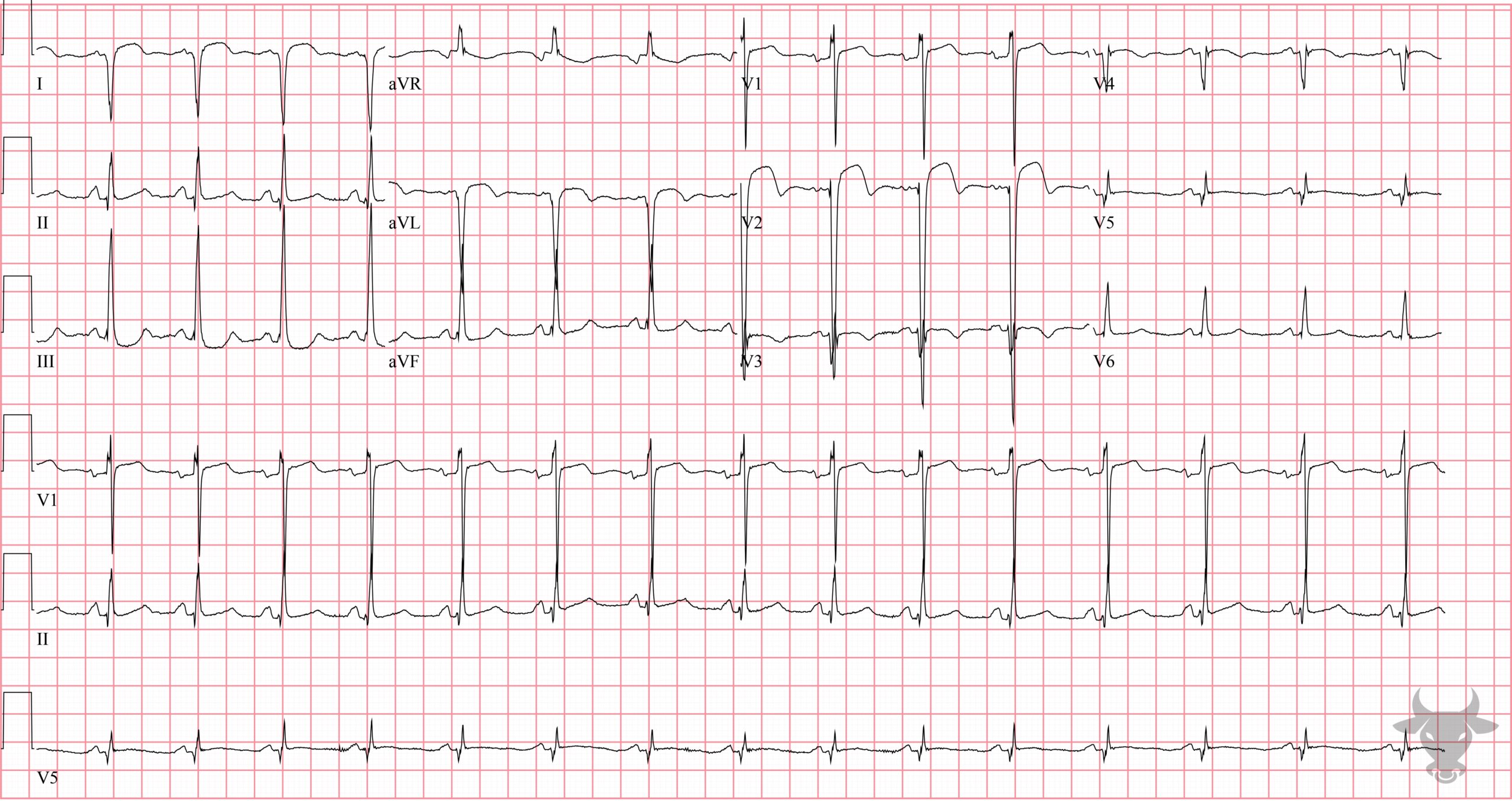
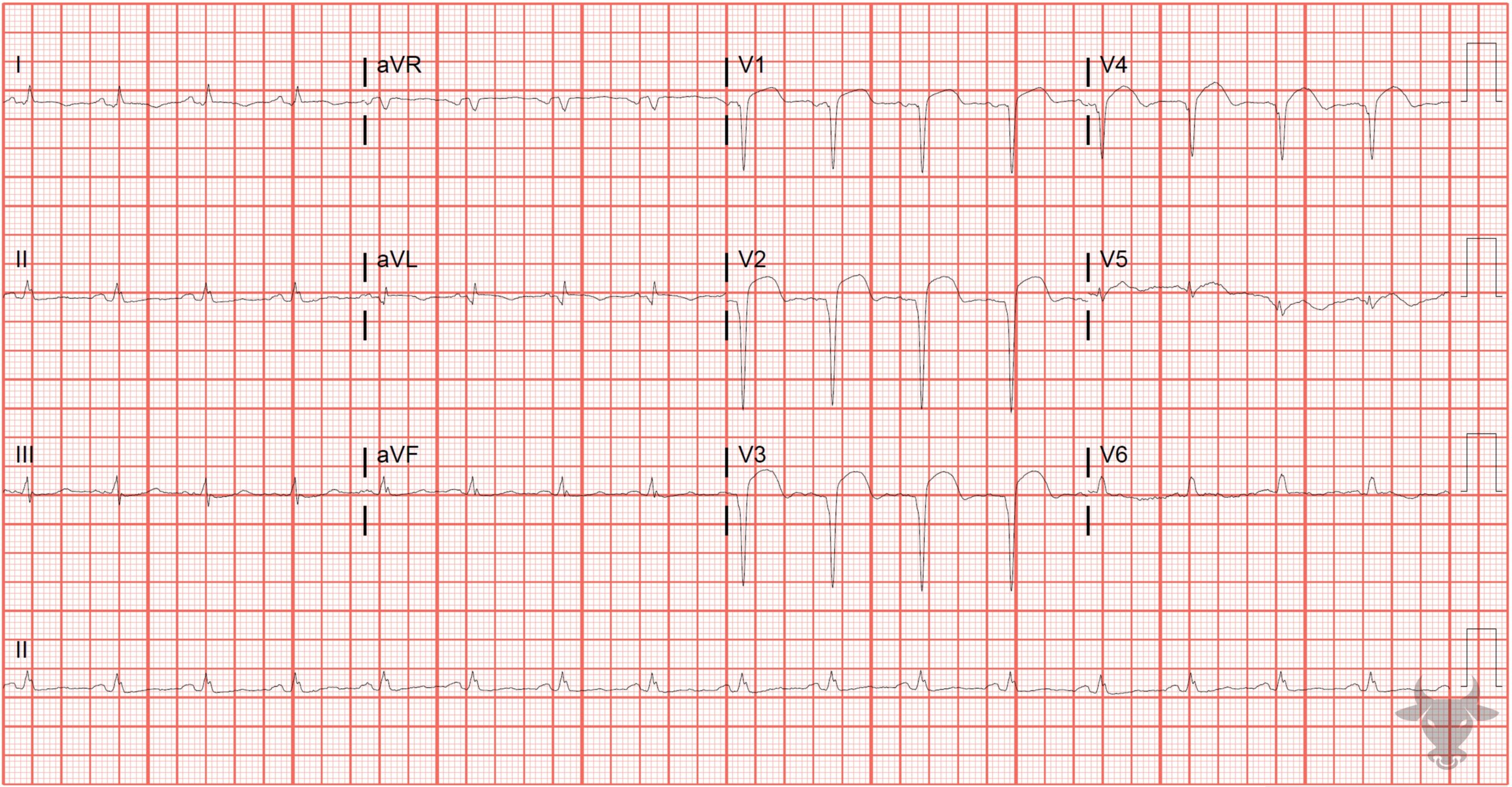
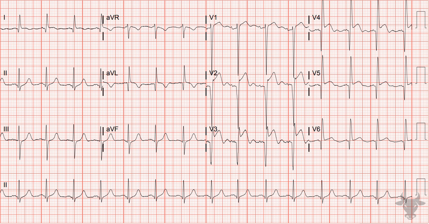
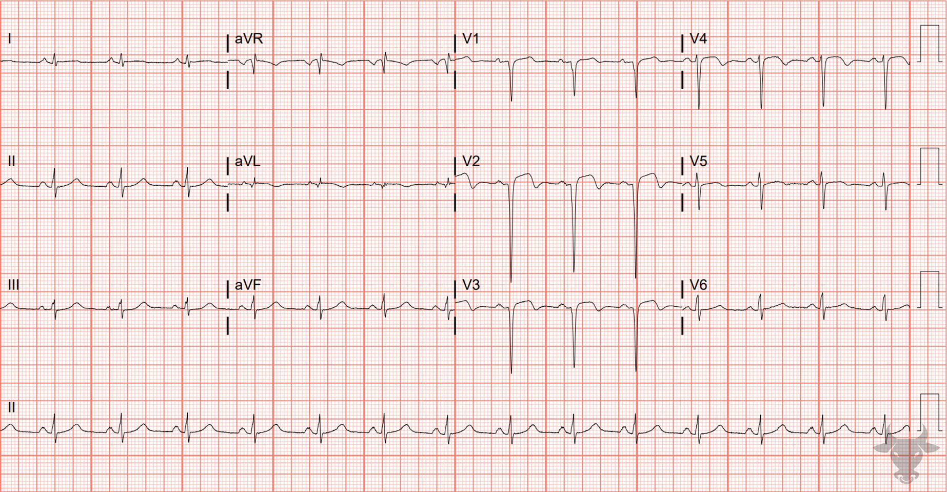
In the age of reperfusion, left ventricular aneurysms have become quite rare, but in the absence of reperfusion, it is a common structural complication of acute myocardial infarction, occurring in 35% to 64% of anterior myocardial infarctions. Left ventricular aneurysm is characterized by echocardiographic ballooning at the apex of the left ventricle or apical akinesis. The usual ECG findings of left ventricular aneurysm include ST elevation that persists more than two weeks after STEMI, deep Q waves, and the absence of reciprocal ST depressions.
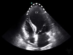
Apical four chamber echocardiogram showing severe balloon-like dilation of the left ventricle (LV) apex (triangles).