Hypokalemia, hypomagnesemia, and hypocalcemia can all cause a prolongation of the QT interval. While hypokalemia and hypomagnesemia both delay the repolarization phase (phase 3) of the of the cardiac action potential (creating wide-based T waves, U waves, or a fusion of both), hypocalcemia prolongs the QT interval by way of extending the plateau phase (phase 2) of the cardiac action potential (lengthening the ST segment but with a normal T wave). The presence of a prolonged QT interval places the patient at risk for Torsade de Pointes.
Hypokalemia

Examples
Hypokalemia
Hypokalemia tends to prolong the QT interval by way of delaying repolarization, creating a broad-based T wave, U waves, and T-U fusions.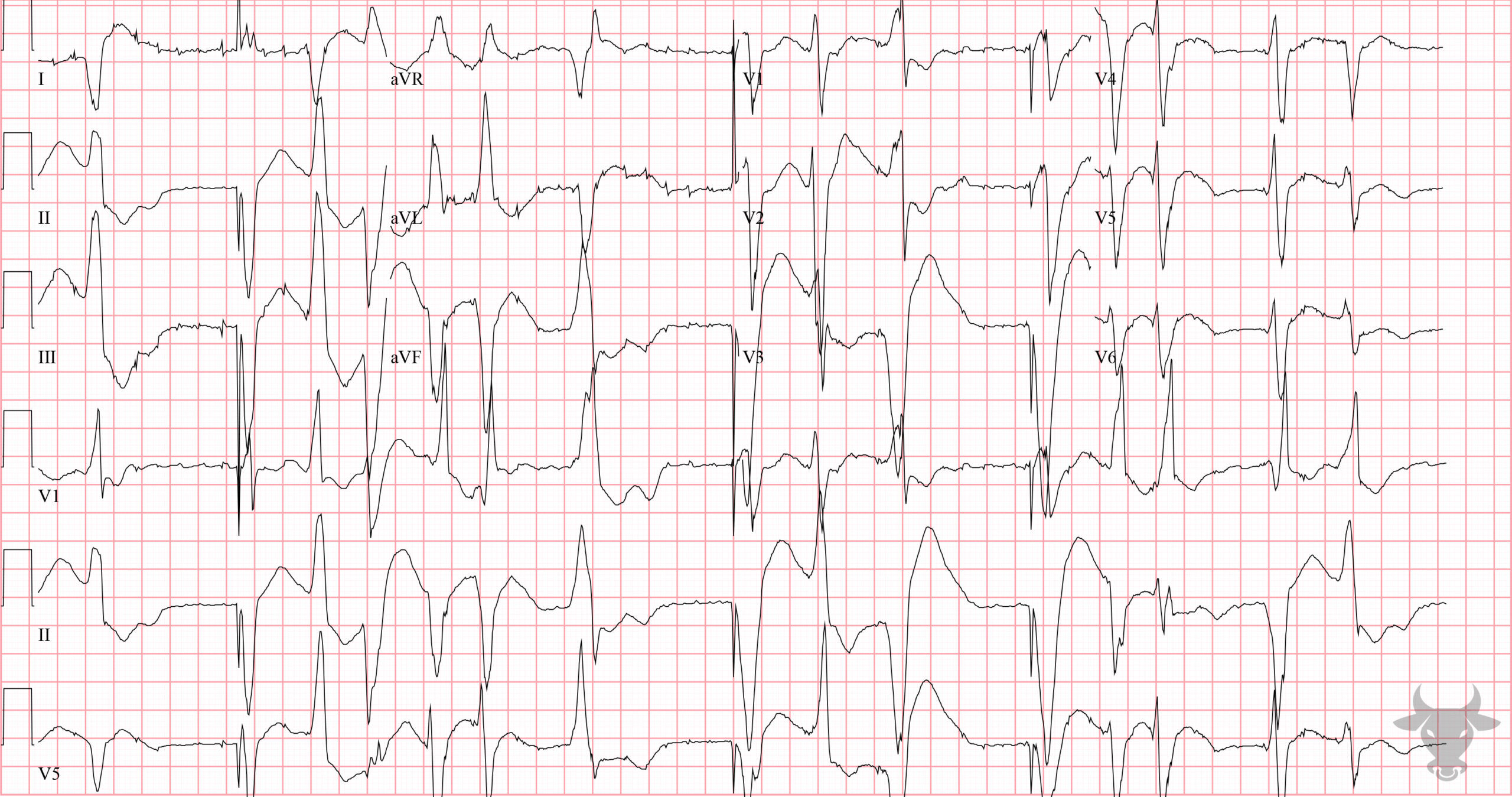
Hypokalemia
Frequent ventricular ectopy places this patient at risk for torsade de pointes.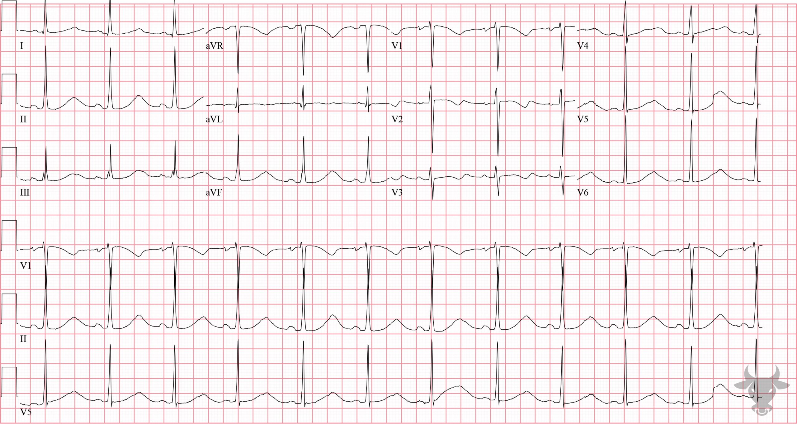
Hypokalemia
There is frequent ventricular ectopy placing this patient at risk for torsade de pointes.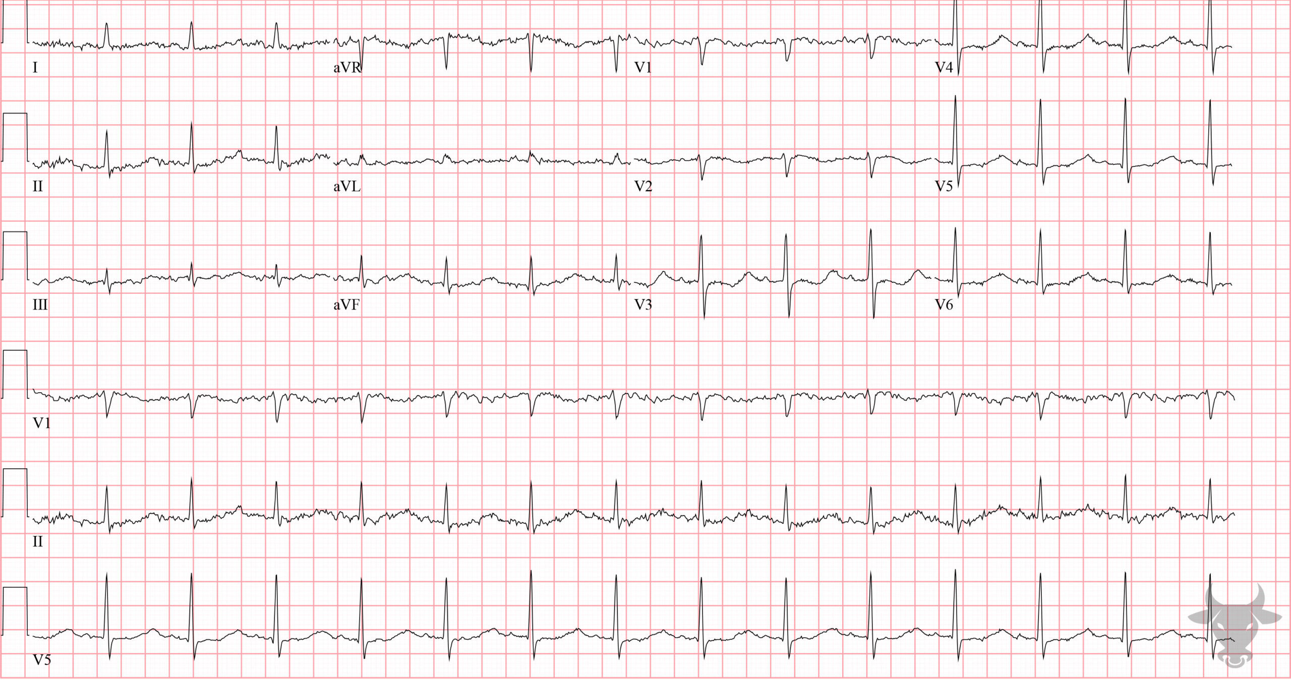
Hypokalemia
This patient had a potassium of 1.5 mEq/L and presented with frequent falls. Notice the prolonged repolarization phase. The downward deflected T wave in the anterior precordial leads fuse with the U wave and create a wavy pattern characteristic of hypokalemia.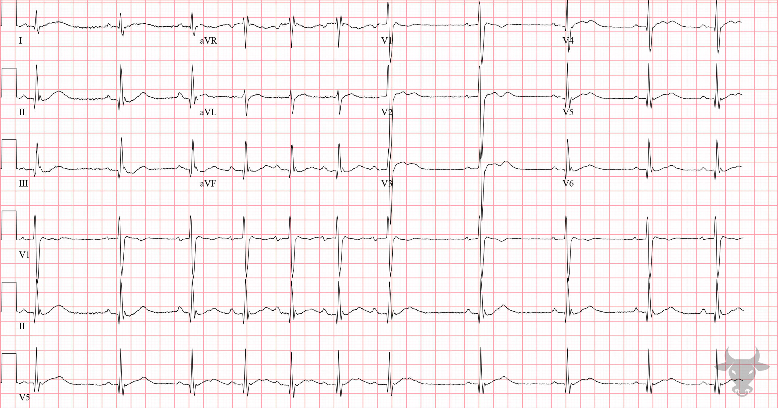
Hypokalemia
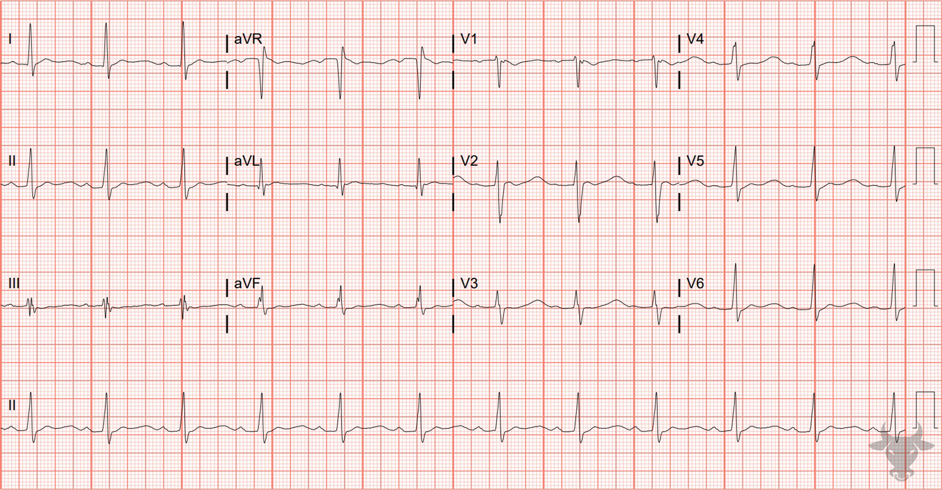
This patient presented with global weakness and was diagnosed with hypokalemic periodic paralysis. The potassium level was 1.9 mEq/L. Note the prolonged QT interval and the U waves.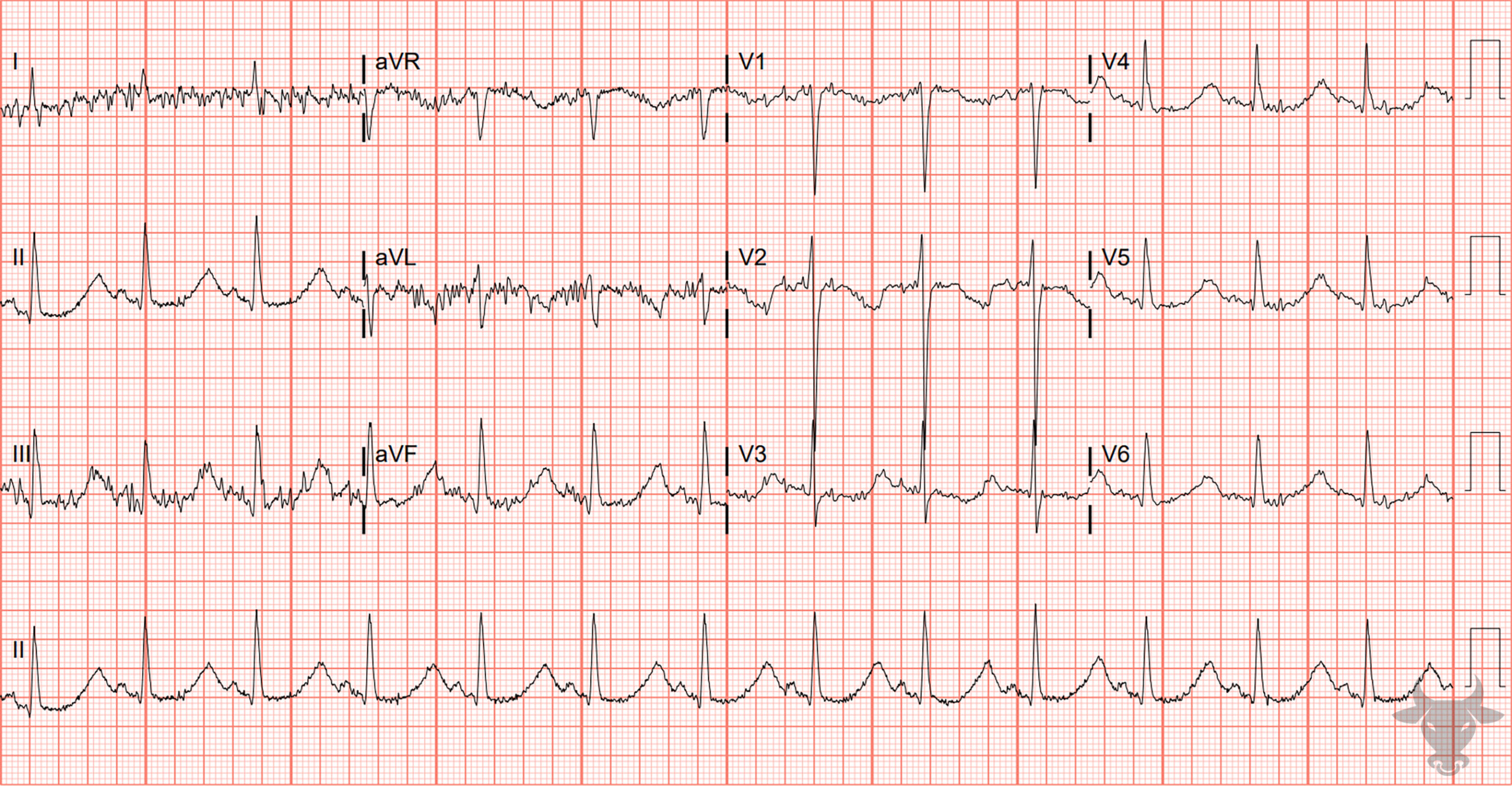
Hypokalemia
While there is quite a bit of baseline artifact, a prolonged QT interval with broad-based T waves is evident. This patient was severely hypokalemic and intermittently went into polymorphic ventricular tachycardia. Overdrive pacing via a temporary transvenous pacer was initiated to avoid further episodes of polymorphic ventricular tachycardia.References
- Diercks DB, Shumaik GM, Harrigan RA, Brady WJ, Chan TC. Electrocardiographic manifestations: Electrolyte abnormalities. Journal of Emergency Medicine. 2004;27(2):153-160.
- Al-Khatib SM, Stevenson WG, Ackerman MJ, et al. 2017 AHA/ACC/HRS Guideline for Management of Patients With Ventricular Arrhythmias and the Prevention of Sudden Cardiac Death. Journal of the American College of Cardiology. 2018;72(14):e91-e220.