Atrial flutter occurs from a reentrant circuit around the tripcuspid valve in the right atrium. It is an organized rhythm that is generally characterized by an atrial rate around 300 bpm, and a heart rate around 150 bpm. It more commonly occurs in a counterclockwise fashion, leading to inverted flutter waves in the inferior leads (II, III, and aVF). Consider 2:1 flutter in any tachycardia with a ventricular rate of 125 to 200 bpm, most commonly close to 150 bpm.
Atrial Flutter

Examples
Atrial Flutter With 2:1 Conduction
This is atrial flutter with 2:1 conduction. Notice the sawtooth pattern in the inferior leads (II, III, and aVF), with inverted flutter waves (i.e., typical atrial flutter).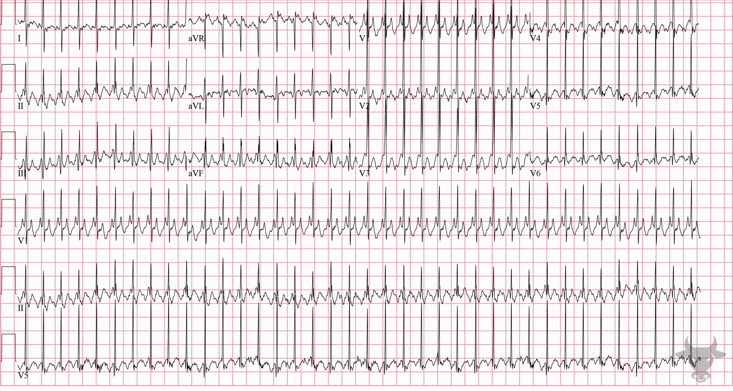
Atrial Flutter With 2:1 Conduction
2:1 atrial flutter in an infant. There is an atypical appearance to P waves in the limb leads (specifically II, III and aVF) where the usual isoelectric baseline is replaced by identical appearing “sawtooth” waves. Pediatric intervals tend to be shorter, and rates faster than in adults (e.g., > 200 beats per minute in this ECG). In the absence of treatment or atrioventricular block, the most common atrial to ventricular response is 2:1.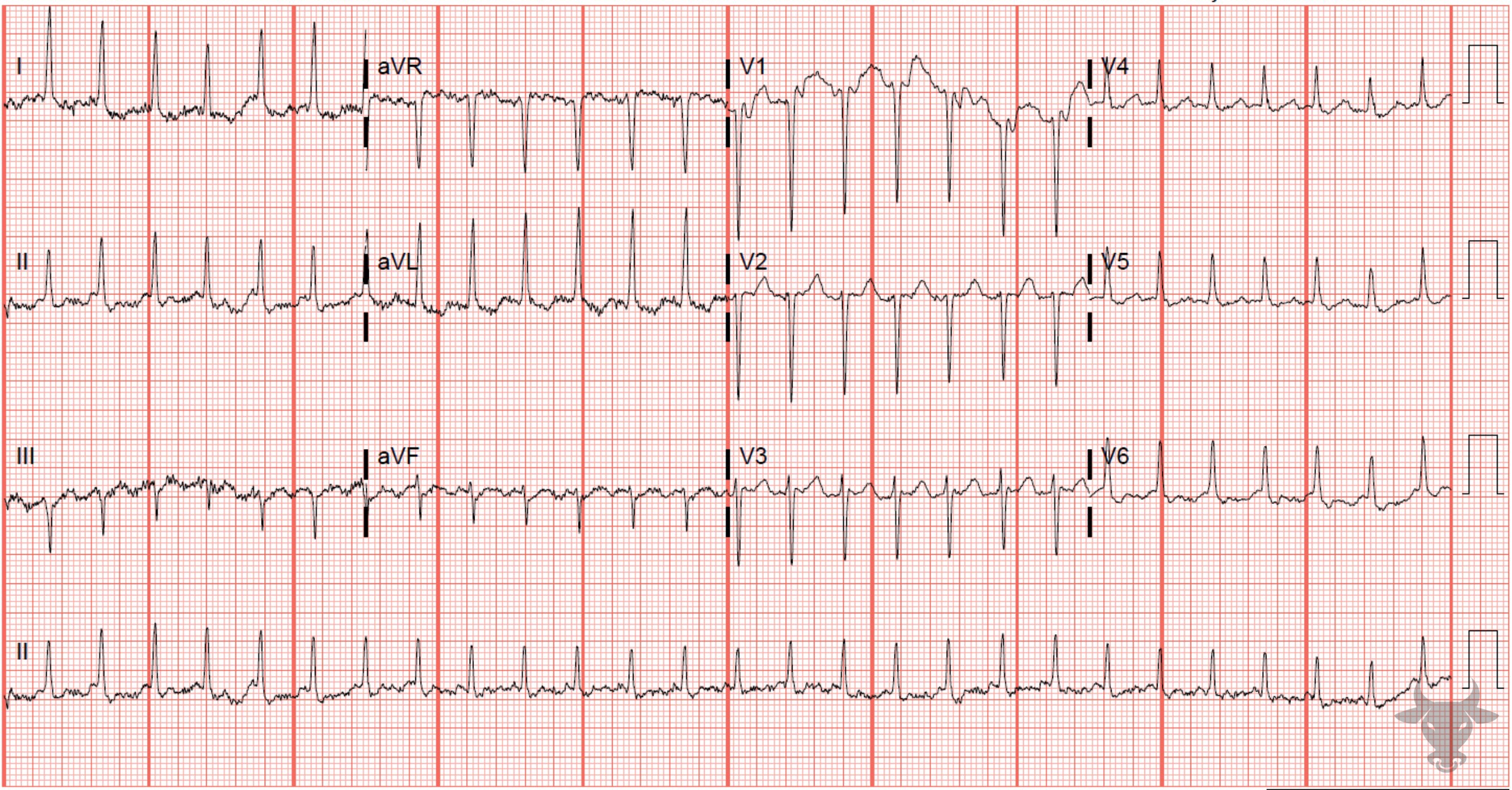
Atrial Flutter With 2:1 Conduction
A subsequent ECG was performed with the Lewis lead, which unmasked the underlying 2:1 conduction. The Lewis lead describe a different positioning of the limb leads in order to better visualize atrial activity.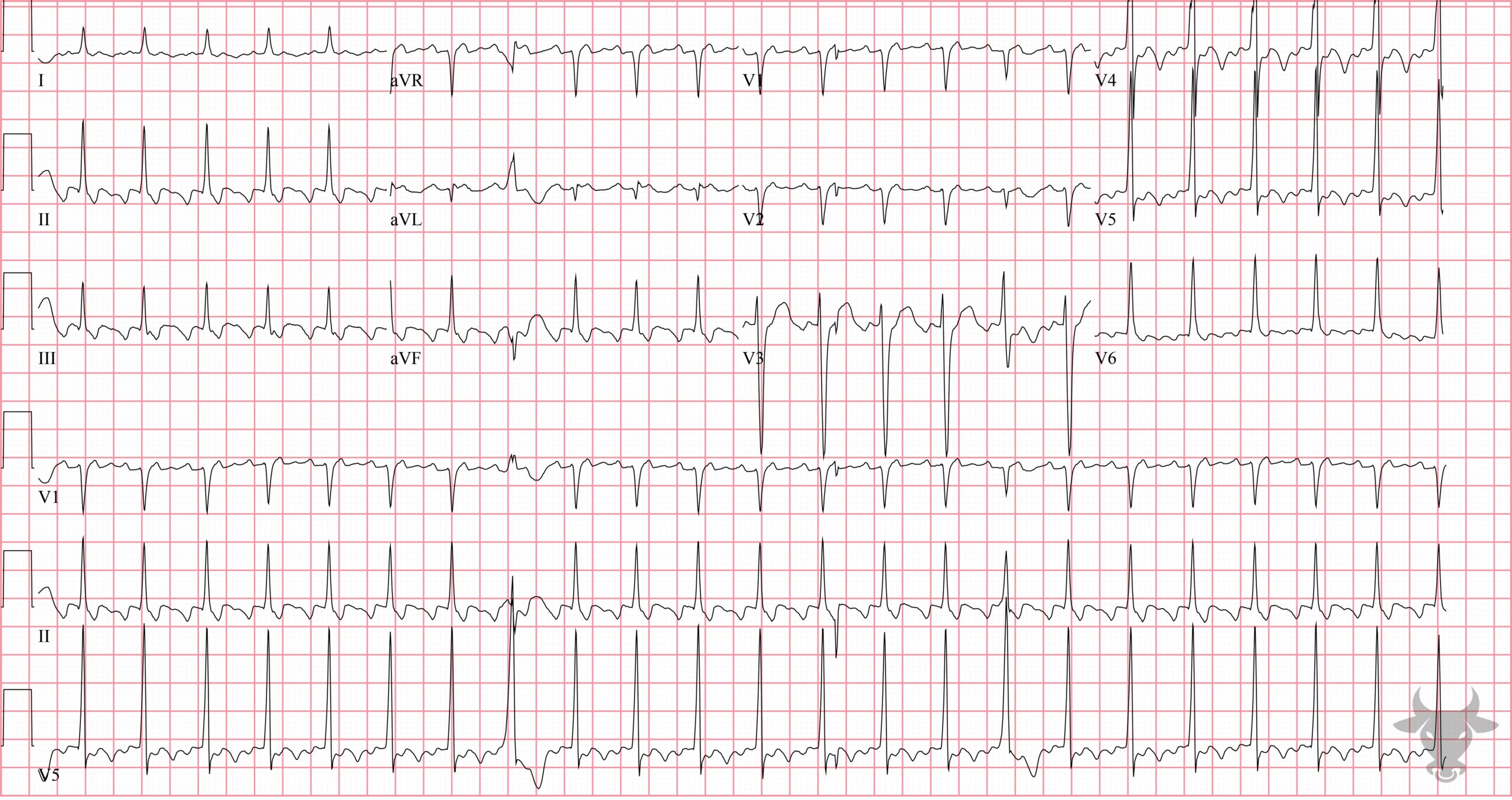
Atrial Flutter With 2:1 Conduction
There is an atypical appearance to P waves in the limb leads (specifically II, III and aVF) where the usual isoelectric baseline is replaced by identical appearing “sawtooth” waves. These atrial flutter waves occur at a rate of approximately 300 beats per minute, complemented by a ventricular rate of 150 beats per minute. In the absence of treatment or atrioventricular block, the most common atrial to ventricular response is 2:1.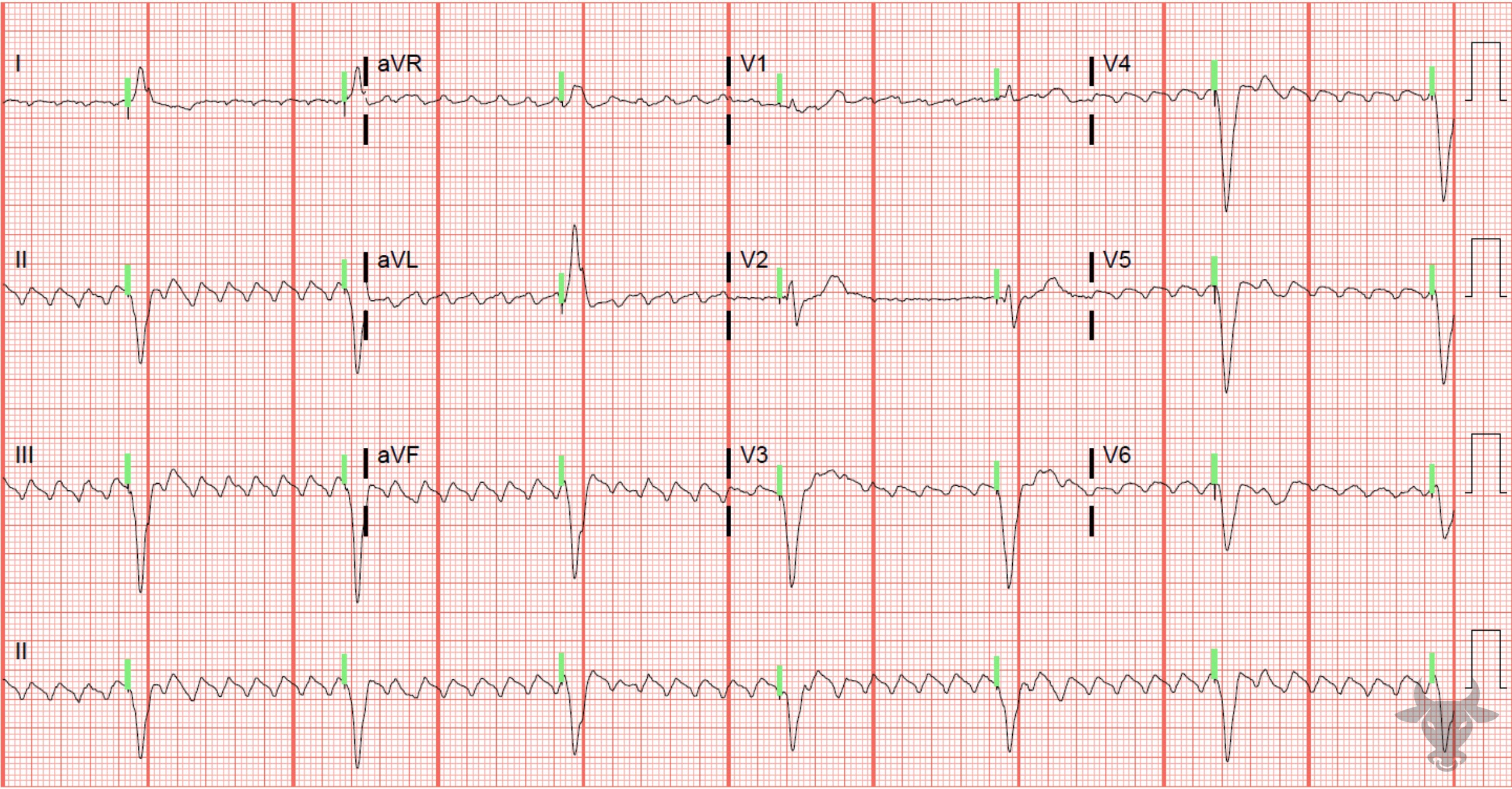
Atrial Flutter
Atrial flutter without native conduction but with paced ventricular activity. The pacemaker threshold was set to 40 beats per minute, and was later increased to 60 beats per minute.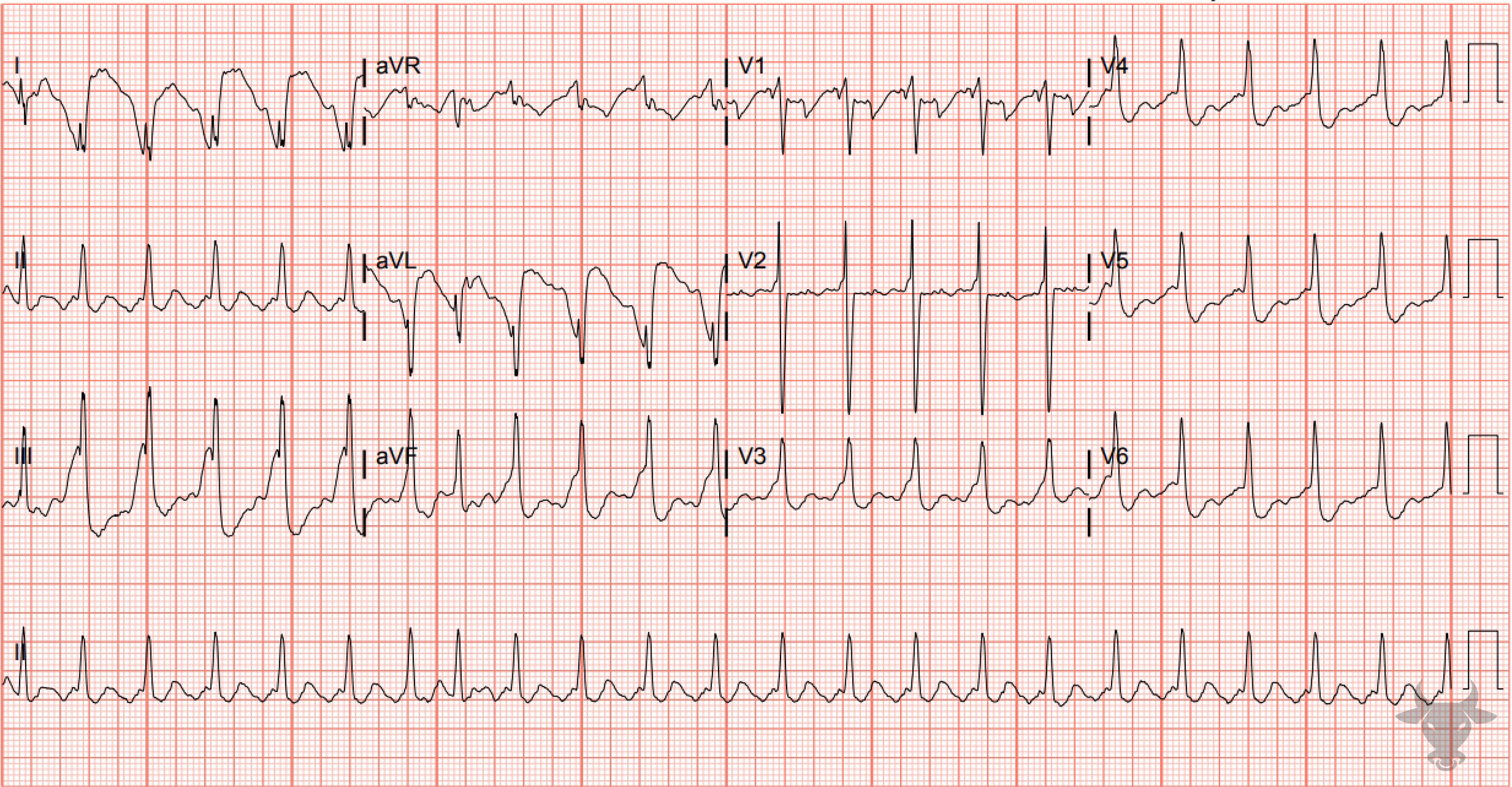
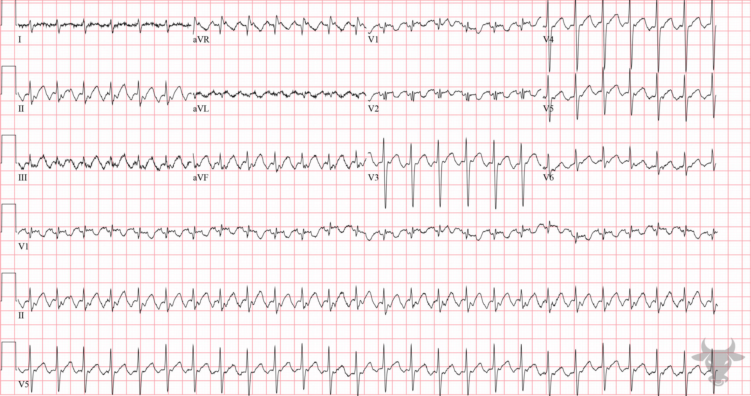
Sodium Channel Toxicity
Atrial flutter with 2:1 conduction. The wide and fragmented QRS, right axis deviation, and tall terminal R wave in aVR are all consistent with sodium channel toxicity; in this case, due to cocaine toxicity.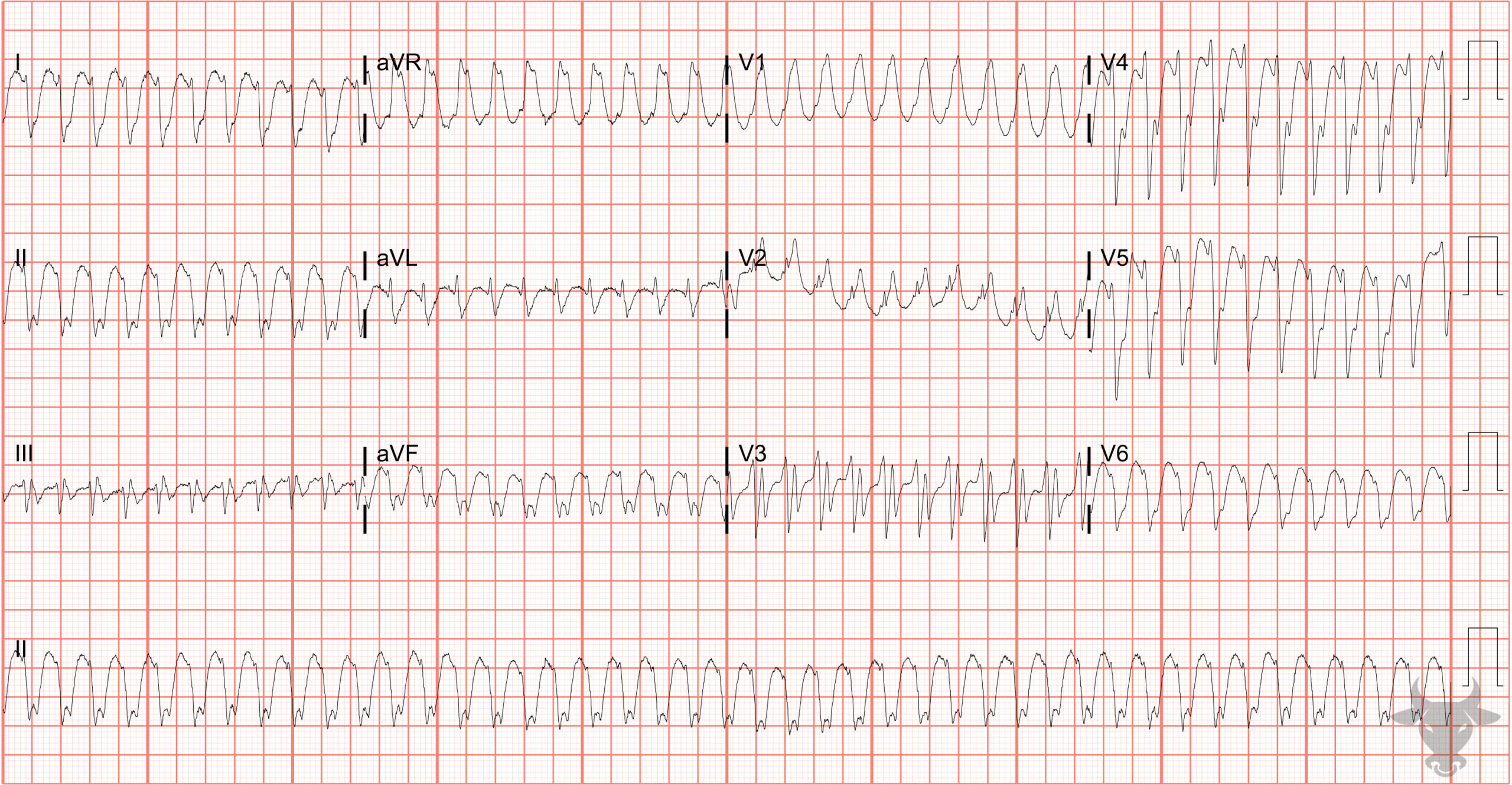
Atrial Flutter With 1:1 Conduction
Here is a wide complex tachycardia with a rate of 260 bpm. While ventricular tachycardia is the most likely diagnosis, the QRS morphology matched prior ECGs in this patient with known right bundle branch and left anterior fascicular blocks suggesting supraventricular tachycardia with aberrancy. Amiodarone was given and the rate cut exactly in half with atrial flutter waves visualized in a pattern of 2:1 conduction, confirming the diagnosis of atrial flutter with 1:1 conduction.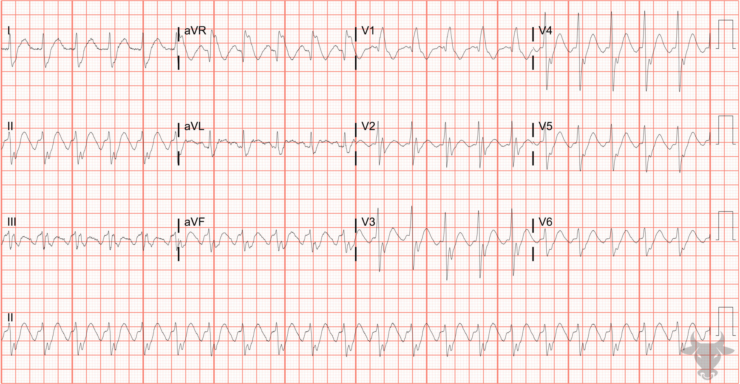
Atrial Flutter With 2:1 Conduction
This patient presented with atrial flutter and 1:1 conduction at a rate of 260 bpm. This ECG was obtained after amiodarone administration slowed the rate to 130 bpm. The patient had known right bundle branch and left anterior fascicular blocks.References
- Link MS. Evaluation and Initial Treatment of Supraventricular Tachycardia. New England Journal of Medicine. 2012;367(15):1438-1448.
- Surawicz B, Knilans TK. Chou’s Electrocardiography in Clinical Practice. 6th ed. Elsevier; 2008.